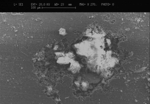
SEM microscopy is a vital part of any metallurgical investigation. SEM imaging can help identify cracks, contaminants, or fractures on the surface of a material, which means that we can understand the behaviour and characteristics of a sample at higher magnification levels than traditional microscopes. Typical features of Scanning Electron Microscopy (SEM Microscopy) that can be viewed are fracture faces of failed parts, or surface conditions of components.
This information is crucial to our customers, as it helps them to understand the surface characteristics of the object under examination, identifying anomalies, defects and elemental composition of the material under scrutiny.
Knowing whether a material is susceptible to specific types of failure, such as fatigue or stress corrosion cracking, or if there are contaminants present at the surface, allows our customers to make informed decisions about the production processes, and if their choice of material is appropriate for its intended application.
In conjunction with SEM microscopy, energy dispersive x-ray spectroscopy (EDS or EDX) is a technique that uses x-rays emitted from the sample during the SEM process to further characterise the composition of the material. EDX, also variously known as EDAX or EDS, EDX microanalysis is the targeted analysis of a feature observed on the scanning electron microscope.